GET STARTED
Milestones in Urologic Education: Ancient Times
Glenn M. Preminger, MD; Russell S. Terry, MD & Ron Rabinowitz, MD
Empirical Beginnings:
Urological Education in Ancient Civilizations
Medical knowledge in ancient times developed as a blend of empirical observations, practical surgical experience, and mysticism. Ancient civilizations recognized the diagnostic value of urine, the complexity of the urinary tract, and the high prevalence of urologic disorders. These factors promoted the evolution of urology as a distinct subspeciality within medicine, a fact which is perhaps best memorialized by Hippocrates’ admonition: “I will not cut for the stone, but I will leave this to be done by practitioners of that craft.” Many of the observations and principles recorded by these ancient forbears, including their educational practices, form the foundation of modern urology as we know it.
Ancient Egypt:
Documenting Urological Knowledge
Ânkh, croix égyptienne
The ankh (Life) is believed to represent the male and female genitalia, the sun coming over the horizon, and the union of heaven and earth.
Wikimedia Commons
In Egypt, medical knowledge was meticulously recorded on papyri, including detailed accounts of many urological conditions. The Ebers Papyrus (circa 16th century BCE) provides insights into medical treatments for urinary retention, bladder stones, and other conditions. The Edwin Smith Papyrus contains detailed case reports of the surgical management of traumatic injuries. Egyptian medical training began in specialized temples called Houses of Life (Per Ankh), where upper-class children learned to read and write. Afterwards, they would apprentice with their family members who were physicians.
Ancient Greece:
The Hippocratic Approach
Hippocrates (circa 460–370 BCE) recognized the diagnostic value of urine and uroscopy examination, stating in the Corpus Hippocraticum that “no other system or organ of the human body gives us so much diagnostic information by its excretion as does the urinary tract.” Hippocrates also astutely noted the high morbidity and mortality risks associated with bladder surgery, and he strongly advised against such procedures except when performed by physicians who were highly experienced in the work—thus laying the groundwork for urology to emerge as a specialized field.
Though the use of human cadavers for education was culturally taboo, the physician scholars Herophilos (335–280 BCE) and Erasistratus (304–250 BCE) made great advancements in anatomical science through the controversial use of dissection and vivisection across the Mediterranean Sea in Alexandria during the 3rd century BCE.
Rome: Galen and Early Anatomy
Roman medical instruments
Donated by the Western Section AUA
Galen of Pergamon (circa 129–216 CE) made significant contributions to anatomy and surgery, drawing heavily from the earlier Greek and Egyptian traditions and adding to them through his pioneer work in comparative anatomy via animal dissection. Similarly limited by cultural views of cadaveric dissection, Galen gained extensive practical surgical experience by treating gladiators, and he emphasized medical education through a combination of theoretical knowledge and practical experience with dissection and direct observation. Galen’s published works spread to the Byzantines in Constantinople, and from there they were translated into Arabic where they helped form the cornerstone of ancient Arabic medicine.
India: Advanced Surgical Instruments
Suśruta
Wellcome Collection
India’s legendary surgeon Suśruta wrote the Samhita (circa 600 BCE), which stands as one of the most comprehensive and advanced surgical texts from the ancient world. This text contained encyclopedic knowledge about anatomy, medical treatments, and surgical procedures and instrumentation. Suśruta described in detail how each of the 125 listed surgical instruments should be manufactured and maintained, including 28 varieties of urethral catheters and sounds.
Unlike his Greek and Roman counterparts, Suśruta was not limited by cultural taboos around dissection of human cadavers for educational purposes. Suśruta therefore emphasized the importance of dissection for the development of anatomical expertise. He also described several different early “simulation models” (fruits, vegetables) which could be adequately used by students to learn and practice basic surgical skills. Suśruta also documented in the Samhita stringent trainee selection criteria, an ethics oath, and an elaborate initiation ritual marking passage into medical apprenticeship.
Focus On: Ancient Egypt & Ancient Greece
Ebers Papyrus
Wellcome Collection
Cast of Thoth, Egyptian Deity of Medicine
Wellcome Collection
Five Men at a Dissection Table, 1482
Wellcome Collection
Hippocrates teaching uroscopy to students, 1488
La Mer des Histories
Focus On: Rome & India
Ancient herbalists and scholars of medicinal lore. Woodcut, 1532
Wellcome Collection
Galen of Pergamon, lithograph, 1815
Wellcome Collection
Images from Suśruta. An English Translation of the Sushruta Samhita
Book donated by Sakti Das, MD
Relief with surgical instruments presented to Imhotep
Image courtesy Sakti Das, MD
Beacons of Light: Urologic Education in the Dark Ages
John Phillips, MD & Dinora Murota
Medieval Urologic Education
Uroscopy Wheel
Epiphanic Medicorum by Ulrich Pindor, 1506, of Nördlingen, Germany, depicting various colors of urine within flasks or ‘matulas’ in Latin.
Wellcome Collection
Medieval education may sound like an oxymoron but there were many examples in the Dark Ages of physicians providing urologic care and teaching methods to do so. Urologic conditions were a great scourge in Medieval times and included urinary and genital skin infections, urethral strictures, and bladder stones. The average lifespan of the medieval European was just 40 years. Those fortunate enough to live longer sought urologic care for the diseases of ‘aging’ including erectile dysfunction (ED), infertility, lower urinary tract symptoms, and urologic cancers.
Urologic education was limited by the availability of textbooks and material. There were some famous medical schools at Salerno, Bologna, Parma, and Paris where students were provided lectures and dissections, but most physicians were trained as apprentices who strictly followed the empiric instructions of their mentors. The Middle Ages was a period of great travel: international commerce and travel carried diseases into cramped, filthy cities and spread plague and pestilence.
Women in Urologic Education
From Chirurgia, c 1180, by Rogerio of Parma, with visual table of contents to provide details of operations for (top left – bottom right) malignant tumors of the breast, mastitis, penile laceration, penile malignancy, an unknown penile condition, and scrotal swelling due to hernia.
Public Domain, British Library, Sloane MS 1977, folio 7v

Monasteries were often a source of protection and urologic care. Hildegard of Bingen, a nun in Germany, wrote great texts on health care including urologic passages on the function of the kidneys (and why there are two), the testicles (and their function in erections), and urinary continence. Hildegard’s work was used for generations to identify salves, diets, and medicines that would be helpful for infertility, ED, kidney stones, and urinary burning.
Trota, an Italian physician near Salerno, was the source of a huge body of medical teachings known as the Trotula. She recognized that women may not have received adequate care from male physicians. The Trotula were designed to address uniquely female urologic disorders like incontinence and prolapse, dysmenorrhea, and obstetric emergencies. Less is known about Trota than about Hildegard, but their teachings were equally important in promoting urologic education and access to care.
Urologic Surgery Education
Urologic surgery was limited by the lack of anesthesia and thorough anatomic knowledge. This did not prevent early surgeons trying their best to relieve urologic suffering. Rogerio of Parma may have been the first surgeon to make a surgical ‘textbook’ with illustrations. His work, the Rogerino, and those of his student Rolando, showed a system-based focus on diseases of the breast, head, face, chest, abdomen, extremities, and genito-urinary system. Rogerio and Rolando’s works describe surgical interventions for hernia, scrotal and penile cancers and wounds, urethral stricture, and bladder stone. Their descriptions of urologic surgeries, written by hand and translated into many contemporary vernacular languages, lack modern accuracy of detail but do show that the medieval mind was already considering how to best transmit surgical knowledge to others.
Uroscopy and Bloodletting
The medieval period was inevitably limited by strongly held beliefs in fable, astrology, and a fuzzy logic about the human place in nature. Physicians followed strict rules about diseases based on seasons and stars, believing that the same forces were responsible for disease. Belief in the four ‘humors’ (espoused by Galen), and that any disease or disorder was due to the imbalance of the humors was held to be law. Examining a person’s urine in a flask, known as uroscopy, was thought to indicate which humors were out of balance. Complicated uroscopy tables were made, forerunners of modern nomograms, that allowed the physician to match a patient’s urine color with conditions and diseases. Bloodletting was such an important basis of health care that it persisted for centuries after the Dark Ages. However, drawings of bloodletting men were popular in medieval textbooks that showed where on the body bloodletting can be done and - almost as important - when in the year or astrological sign. Urologic conditions such as kidney or urinary stones, bladder difficulties, or ED were addressed through bloodletting from the penile or scrotal veins.
Physician lecturing to students about uroscopy; he points to a flask held by an assistant. Heliotype.
Wellcome Collection

Page from the Trotula
Europeana
Operation for scrotal hernia
Biblioteca Casanatense
Bloodletting man
Circa 1410, illustration showing the preferred sites for venous bleeding to relieve indicated symptoms.
Library of Congress
Cartography to Surgery: The History of the Development of the Urologic Surgical Atlas
Sutchin Patel, MD; Ron Rabinowitz, MD & Friedrich Moll, MD
Defining the Atlas
Theatrum Orbis Terrarum
Wikimedia
Atlas, in Greek mythology, was a Titan condemned by Zeus to hold up the heavens for eternity. The Flemish geographer Gerard Mercator (1512-1594) was the first to use the term ‘Atlas’ as the title of a bound collection of maps. His Atlas Sive Cosmographicae Meditationes de Fabrica Mundi ed Fabricati Figura (Atlas or cosmographical meditations upon the creation of the universe and the universe as created) was first published the year after his death. This defined the atlas as a collection of maps, bound in one or more volumes, whether or not augmented with text. Although individual maps had been drawn for millennia, it was the Flemish cosmographer Abraham Ortel (Ortelius) (1527-1598) who is credited with producing the first atlas. In 1570, he published Theatrum Orbis Terrarum (Theatre of the World), a book of a uniform collection of similar sized map sheets bound together with sustaining text. There were four versions of the first edition, and, at the time, it was the most expensive book ever printed. It continued to be published for 14 years after Ortelius’s death, totaling 7,300 copies in 31 editions.
The First Surgical Atlases
Bourgery & Jacob, “Traite complet de l’anatomie de l’homme”, 1854
Wellcome Collection
One of the first surgical atlases published was Traité complet de l’anatomie de l’homme (The Complete Atlas of Human Anatomy and Surgery) by Jean-Baptiste Marc Bourgery (1797-1849), a French physician and anatomist. Bourgery began work on his atlas in 1830 with illustrator Nicolas Henri Jacob. The first volumes were published in 1831, but the finished work took nearly 20 years to complete (1854).
Dr. Robert M. Zollinger (1903-1992), Dr. Elliot C. Cutler, (1888-1947), and illustrator Mildred Codding (1902-1991) first published Atlas of Surgical Operations in 1939. The target audience of the first atlas was not only practicing surgeons and trainees, but also general practitioners who were still performing operations, despite the establishment of the American Board of Surgery certification in 1937. The clarity of Codding’s illustrations and the purposeful use of a distinctly large format with text facing the drawings made the book easy to read.
The First Urology Textbooks
Young, H. H., Davis, D. M., & Johnson, F. P. (1926). Young’s Practice of Urology: based on a study of 12,500 cases. Saunders
William P. Didusch Center for Urologic History
Many of the first urology textbooks had sections describing early urological surgery. Young’s Practice of Urology (1926) by Hugh Hampton Young and David M. Davis had chapters on the operation of the kidneys, ureter, bladder, prostate, seminal vesicles, scrotum and scrotal contents. urethra, and penis (Volume 2, Chapters 14-21). The text had over 1,000 illustrations by William P. Didusch. Short surgical handbooks such as Surgical Urology: Handbook of Operative Surgery by R.H. Flocks and David Culp (1954) were the next evolution of educational tools used to help teach urological operations.
Frank Hinman Jr.’s Atlas of Urologic Surgery (1989) and Atlas of Pediatric Urologic Surgery (1994), both illustrated by Paul Stempen, served to become some of the most widely used atlases in urology. In the preface to the first edition of his atlas, Hinman would write, “soon after completing residency training I began to record with sketches and brief notations the techniques I was learning and teaching the residents…I intended to put together a “how-to” atlas, modeled after that of my fourth-year surgery teachers, Cutler and Zollinger.” In describing the construction of his atlas he wrote, “I reviewed my sketches and post operative notes made over the last 35 years. Current and classic publications were then consulted to be sure that each important step of every operation was covered…I described the operation step by step, just as I would tell you how to do it at the operating table, ‘Cut here, suture here.’ I hope the user won’t take offense at this approach.”
Evolving Medical Technology
Dr. Frank Hinman Jr. and a page from his Atlas of Urologic Surgery.
Hinman, Frank, Jr., ed., (1989) Atlas of Urologic Surgery, W.B. Saunders Company, Philadelphia, PA.
William P. Didusch Center for Urologic History
With changes in medical technology, surgical atlases have evolved to include atlases of cystoscopy and atlases of laparoscopic and robotic surgery. The next evolution in surgical teaching occurred with the creation of video libraries of surgeries often accompanied by narration.
Surgical atlases have served as teaching tools helping generations of surgeons prepare for the operation at hand. The evolution of these atlases reflects the dynamic relationship between technology, art and medicine.
The Dead Teaching the Living: A History of Corpses in Surgical Education
Connor Hartzell, MD & Jennifer Gordetsky, MD
Human Anatomy
The modern study of human anatomy originated in the 11th century when, after centuries of neglect, European medicine refocused on the doctrines of common sense and observation. The discipline of anatomy’s founding principles permeate to the present: studying the normal human body could help teach normal function and studying the abnormal could help understand pathology. The earliest recorded dissections of this era were the autopsies of homicide victims. This led to the formation of anatomy as a core medical science.
Dissection in Education
Classroom with a group of students and three cadavers, c. 1915
National Library of Medicine

During the Renaissance, interest in the discipline grew. Grave-robbing and other forms of body snatching by anatomists and their students was common. Dissections were performed primarily at teaching institutions such as Leyden University in Netherlands. The studies of anatomy and experimental medicine led to major discoveries including the circulation of blood (William Harvey) and improvement of surgical techniques. Throughout the 1700 and 1800s, medicine in Europe continued to advance with knowledge acquired through anatomic study. This was brought to the New World with the openings of the first American medical schools. Dr. William Welch, a pathologist at Johns Hopkins in the mid-1800s, gave autopsy seminars that not only became part of the core curriculum at his institution, but influenced medical education standards nationwide.
In the early 1900s, medical science was advancing rapidly, and the demand for cadavers to train physicians increased. Grave-robbing was outlawed in the late 1800s, so training cadavers were often unclaimed bodies from prisons and other state institutions. Body donation became more acceptable mid-century, and today, most cadavers for American medical education are acquired through donation with consent.
Dissection in Controversy
Although physicians came to use dissection as a critical tool in medical and surgical education, the acquisition and dissection of human remains met strong resistance throughout history. In the 1300s, the Catholic Church strongly opposed human dissection, although this did not stop these procedures from occurring in universities. In the New World, William Shippen (1712-1801), Professor of Anatomy at Philadelphia’s College of Physick, was accused of grave robbing and attacked by a mob for showing specimens from human dissections. It was only with the introduction of embalming techniques and the rise of the funeral industry in the late 1800s that the public fully accepted the practice of cadaver dissection.
Cadavers in the Law
Robust legal doctrine establishing the rights and uses of so-called “anatomical gifts” (organs and cadavers) arrived with the passing of the Uniform Anatomical Gift Act (UAGA) of 1968. It controversially did not address the legality of using unclaimed bodies and paying for bodies for purposes other than organ transplantation/therapy, which it explicitly outlawed. To this day, major disagreements regarding the use of unclaimed/purchased bodies in medical education persist.
In today’s surgical training programs, cadavers remain crucial to honing routine skills and practicing rarely used techniques. Simulations using fresh cadavers have improved urology residents’ confidence and skills in robotic surgeries. Looking to the future, new technologies such as virtual reality may augment traditional cadaver-based anatomic education. The “hands-on” value of a body, however, will never disappear.
Dissection Lessons Throughout Time
Bartolomeo Eustachi, Tabulae Anatomicae, 1714
Anatomical theater dissection; a surgeon examines the cadaver; several men observing.
National Library of Medicine
Anatomical Demonstration
Dissection of cadaver for demonstration purposes
National Library of Medicine
Dissecting room at Chattanooga Medical College
Prof. Ellis, Dr. Woolford, and Class in Anatomy
National Library of Medicine
Flayed cadaver holding his skin
Juan Valverde de Amusco, Anatomia del Corpo Humano, 1559
National Library of Medicine
Prematurely interred person rises after being exhumed by a terrified grave robber. Jacques Benigne Winslow, Uncertainty of the Signs of Death, 1746
National Library of Medicine

Genitourinary collecting system from body donated to Vanderbilt University Medical Center
Jennifer Gordetsky, MD

Vanderbilt University School of Medicine Gross Anatomy Lab for medical students
Anna Edmondson, MD

From Early Modern Apprentices to Modern Medical Students
David Bloom, MD & Barbara Chubak, MD
Early Modern Medical Learning
The early modern period (called the Renaissance in Italy) featured a revival of Greco-Roman humanism and interest in the works of Galen. Physicians who learned medicine in universities grappled with the tension between theory and practice: Galenic medicine encouraged a patient-centered consideration of disease etiology, but lectures and books for medical practice (called Practica) enforced rote memorization of symptoms and their treatment. Surgery was generally taught by apprenticeship, organized through local guilds or colleges of surgeons and barber-surgeons.
Fig 2. Tools used for the proprietary Colot surgery for bladder stone, from Traite de l’Operation de la Taille (1727). The Colot family of French barber-surgeon lithotomists developed a technique for lateral perineal lithotomy in the mid-1500s.
Wikimedia
Pioneering physician Andreas Vesalius established a detailed knowledge of anatomy as fundamental to medicine as well as surgery and brought the Scientific Revolution to health care by arguing that medical knowledge should be generated empirically. However, his perception was skewed by Galenic dogma, as exemplified by his genital dissections that reinforced a perception that the female reproductive parts are an inversion of the male (Fig 1). Surgeons, who favored craft tradition over Latin literature, made readier progress through empiricism, but often kept it secret to maintain a competitive advantage in the medical marketplace (Fig 2).
Education in the Enlightenment
In the 17th and 18th century, medicine evolved away from Classical humoralism, incorporating chemistry and physics to understand the body as a mechanical device composed of interconnected and manipulable parts. This body concept, along with the Enlightenment-era enthusiasm for hands-on experiential learning, further narrowed the divide between physicians and surgeons. The urban accretion of infectious disease and other illnesses prompted the reform of existing hospitals from places of shelter into spaces for health care, and new medical hospitals were sponsored by governments and private philanthropy to accommodate the sick-poor in both the old and new worlds (Fig 3).
Hospitals became schools for student physicians and surgeons alike, who learned by caring for living patients and dissecting the dead. In the 19th century, education followed the “Paris Medicine” model, in which detailed observation of live patients was followed by autopsy to identify the lesions corresponding to their pre-mortem presentation and statistical analysis to determine treatment efficacy. The pursuit of clinical research and experimentation became woven into the fabric of medical education, and American doctors who traveled abroad brought this influence home to their own students.
Learning in the Long 19th Century
Fig 4. Newspaper clipping featuring story about abuse in medical lecture at Pennsylvania Hospital.
Drexel University Archives
By the later 1800s, medical education took place in schools of various types. Germany and the Austro-Hungarian Empire had university-based medical schools, which encouraged interdisciplinary collaboration, specialization of expertise, and the inclusion of laboratory-bench research. In France and Britain, schools were affiliated with hospitals and more clinically oriented in their research and mission. In America, stand-alone proprietary schools varied in quality of teaching, value for tuition money, and ideological orientation, as regulatory requirements for medical practice differed between states, and “regular” medicine competed with homeopathy and other popular medical sects for market share and professional authority (Fig 4).
Americans exposed to European learning inculcated the values of scientific medicine and specialist expertise into American universities and hospitals. Johns Hopkins University, established in 1876, mimicked the German model, which was held up as the ideal form of medical education by Abraham Flexner in his revolutionary report of 1910 that led to the closure of over half of American medical schools in following years. Interested American surgeons embraced specialization, forming the American Association of Genitourinary Surgeons in 1887. From 1890-1892, this organization was renamed the American Association of Andrology and Syphilology, reflecting the field’s foundation in venereal disease, but the stigma of this connection and interest in a broader scope of practice led to a return to its original name. Specialty nomenclature evolved further at the turn of the 20th century, when the older term “genito-urinary surgery” was largely replaced by “urology.”
A woman distilling medication at home, from the frontispiece of “The Accomplished Ladies Rich Closet of Rarities”, 1691
Library of Congress

Fig 1. Image of the uterus as inverted penis, from Vesalius, De Humani Corporis Fabrica
Wellcome Collection
Fig 3. Pennsylvania Hospital
1832, as illustrated by John Caspar Wild. This was the American colonies’ first general medical hospital, established in 1751 to provide care for sick-poor of Philadelphia.
Library of Congress
Fig 5. Dr. Ramon Guiteras, 1880-1890
William P. Didusch Center for Urologic History
Education in Wax
Friedrich Moll, MD
Models in Education
Heart model of an alcoholic human heart, c, 1903. Molded in elastin, painted
Karl Henning, Bohusläns Museum, Sweden
Written in 1552 and printed in 1714, the Tabulae anatomicae of Bartolomeo Eustachi prompted Pope Benedict XIV (1675-1758) to create a Museum of Anatomy in Bologna for which sculptor Ercole Lelli (1702-1766) constructed eight anatomic models of the whole human body. Since the 18th century, it was known that learning from a three-dimensional model was better than learning from a drawing or written text. Previously, only dried specimens or bones were used in teaching.
From Cadavers to Models
Felice Fontana (1730-1805), founder of the Florentine Museum of Physics and Natural History, later made cadaveric specimens into wax models for teaching anatomy to physicians and the general public. This combined several traditions: medical illustration, the modelling of religious icons in wax and the production of colored wax busts.
There was a close collaboration between anatomists, who performed careful dissections following the drawings of anatomical treatises, and modelers who then produced the wax specimens. For obstetrics, teaching aids were made from terracotta, leather or clay.
Wax Models
Wax, however, was easier to sculpt and also cheaper. Wax had been used by ancient Egyptians, Greeks, Etruscans, and Romans to create religious figurines (votives). The moulage was made from white Smyrna or Venice wax mixed with Chinese or plant waxes, mastic, tallow, turpentine, and fats to increase the melting point and elasticity. Once melted, the wax was mixed with finely ground and pre-filtered pigments of body-part-specific color dissolved in turpentine. Layers of wax were then poured into a plaster cast previously moistened with warm water and soft soap to facilitate the detachment of the cured wax.
Muscle Man wax figure, Felice Fontana, 1767/1800
Vienna, Josephinum, Collections of the Medical University of Vienna

Later, Jules Baretta (1834–1923) made more than 2000 wax models in Paris’s Hôpital Saint-Louis, a clinic focusing on disorders of the skin and venereal diseases. In the late 1800s, this museum became the most popular teaching institution for physicians coming from the United States to Europe to specialize.
Models in Medical Specialties
In the early 1800s, wax models and moulages were used in the new medical specialties, such as dermatology or urology. The moulage passed from local use to international acceptance as medical specialties and scientific communication increased. By the 1970s, moulages lost their pre-eminent position as teaching and visual aids to depict dermatological and venereal diseases.
The modern plastination technique (infiltration and embedding of the organs by polymerizing resins) represents a continuation of the wax modeling tradition.
Anatomical Exhibits in Palazza Poggi, Bologna
Wikimedia Commons

Moulage of a gumma in syphilis for training students, University of Tübingen
Wikimedia Commons

Man holding his skin from Body Worlds traveling exhibition
Swamibu

Models illustrating the birth process in Palazza Poggi, Bologna
Wikimedia Commons

Urologic Organizations Standardize Training
Linda Dairiki Shortliffe, MD
Worst to Best: Creating Public Trust
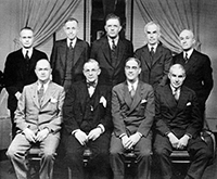
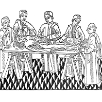
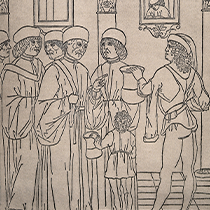
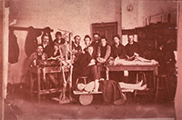
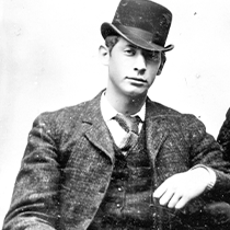
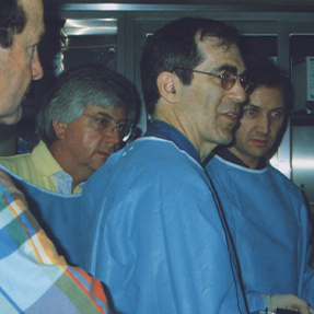
Fig 1. Founding Trustees of the American Board of Urology
The American Urological Association Centennial History
1902-2002, Volume II, pp. 745-756.
In the early part of the 20th century after the Flexner Report (1910), “American medical education [began to evolve] from the worst in industrialized civilization to the very best … the marvel of the industrial world.”
While the Association of American Medical Colleges (AAMC) reformed medical schools, U.S. medical practice lagged in medical knowledge of the time. Postgraduate medical training varied in content, supervision, and duration from weeks to years. Recognizing an oversight gap in postgraduate training, a physician-led group with members of four early boards (Dermatology, Obstetrics and Gynecology, Otolaryngology, Ophthalmology) met to consider a national system to improve training standards. In 1933, this group became the American Board of Medical Specialties (ABMS).
Winds of organization blew within urology as well, and in 1932, the AUA Executive Committee received a report from the Membership Committee noting “…a very intensive movement is going on throughout the country as to the standardization of specialists….” and around the same time, the American Association of Genito-Urinary Surgeons (AAGUS) appointed a committee of three to study specialization in urology. In 1933, the AMA Section on Urology studied qualifications for specialization, and representatives from AUA, AAGUS, and AMA discussed forming a board of Urology: “1) for the protection of patients from unprepared uncertified specialists in urology [and], 2) to raise the standards of education in the special field of urology…”
The next year, the American Board of Urology (ABU) was founded as an autonomous organization with nine members, three from each of the three parent organizations: AUA, AAGUS, AMA. Currently, the ABU has 12 trustees representing 6 urologic organizations. A larger joint AUA-ABU Examination Committee designs the objective written tests. The ABU initiated credentialing: a process to include 1) graduation from an approved medical school, 2) an approved internship and a defined period of residency training or practice or an alternative of preceptorship training, 3) case reports, 4) letters for peer review to assess ethical behavior, and 5) certification consisting of qualifying and certifying examinations. The examinations evolved from freeform to standardized objective written tests, including pathology and radiology, and an oral examination with standard questions. Perhaps most importantly, “unacceptable or illegal behavior” risks revocation of certification.
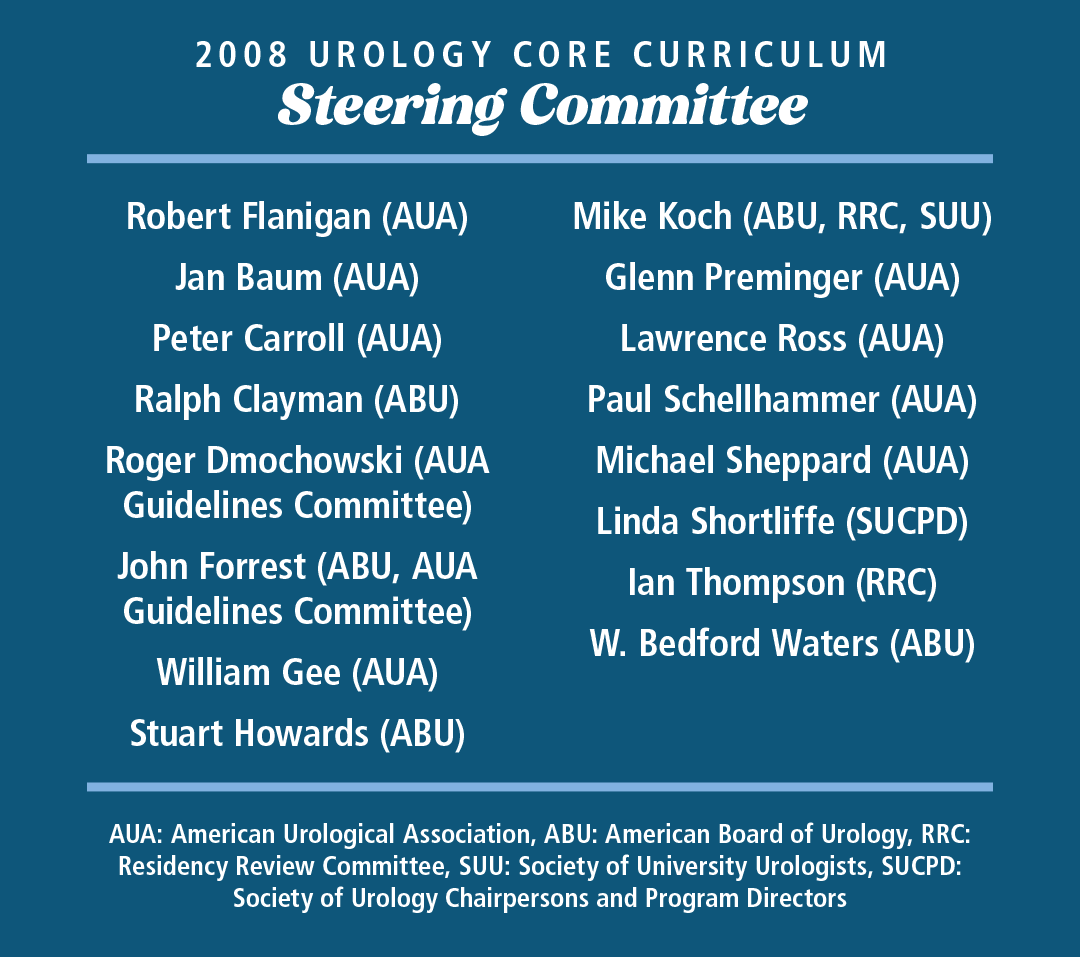
The postgraduate training process (residencies) lacked oversight, however. In 1972, cooperating organizations formed the Liaison Committee for Graduate Medical Education (LCGME, later becoming the Accreditation Council for Graduate Medical Education, ACGME) to accredit specialty residencies with Residency Review Committees in specialties (RRC, Urology). In 2007, a standardized core curriculum was developed after a group of stakeholders - representatives of the AUA, ABU, RRC, AACU, and ACGME - met to discuss the future of Urologic Residency Training.
Preserving Public Trust:
Organizational Accountabilities
Forming the ABU as an autonomous certifying organization was part of assuring the public that a urologist fulfilled credentialing requirements and was certified by examination. With rapid advances, medical knowledge is a moving target, and the adoption of new knowledge by practitioners varies from early adoption to lack of adoption. New technologies enabled the tracking of medical outcomes, and the federal government started tracking physicians with the National Practitioner’s Databank (1986) and the HIPAA Data Bank (1996). However, an Institute of Medicine Report: To Err is Human (1999) exposed medical errors from outmoded knowledge, tests, and management. These events prompted progress in two areas: 1) measures to ensure continuous education and 2) increased sub-specialization.
The ABMS adopted “recertification” for its member boards; the ABU adopted voluntary (1981) and then mandatory (1995) recertification with time-limited certificates to confirm evidence of continuing education. In 2006, this ABU process evolved to “maintenance of certification” (MOC), including a pass-fail examination to complete the recertification process. After an ABU Town Hall in 2017, a program to assess longitudinal learning with continued urologic certification (CUC) without certificate expiration was developed.
With the growth of more diverse and subspecialized urologic practices, post-residency “fellowships” sprouted. Since their scope and requirements differed, training standardization followed different models. In 1989, pediatric urology submitted to the ACGME “Proposed Special Requirements for Programs in Pediatric Urology” with the ABU supporting accreditation without certification. In 2004, the ABMS approved Pediatric Urology as a subspecialty, and in 2006, the ABU approved a Pediatric Urology certification. In 2011, the ABMS approved certification for female pelvic medicine and surgery (Urogynecology and Reconstructive Pelvic Surgery) after a joint ABU and ABOG (American Board of Obstetrics and Gynecology) application.
With changes in medicine and practice, the ABU and other accountable organizations were established, often with controversy, to operate autonomously. While these organizations face future challenges from medical information spread via social media often unpatrolled for misinformation, commercialism, abuse, artificial intelligence, and goals of private equity, the mission is the same.

1847

American Medical Association (AMA)

1876

Association of American Medical Colleges (AAMC)

1902

American Urological Association (AUA)

1910

Flexner Report

1933

American Board of Medical Specialties (ABMS)

1934

American Board of Urology (ABU)

1972

Liaison Committee on Graduate Medical Education (LCGME) and Urology Residency Review Committee (RRC)

1981

Accreditation Council for Graduate Medical Education (ACGME, RRC Urology)

1986

National Practitioner Data Bank

1995

ABU Mandatory recertification

1996

Healthcare Integrity and Protection Data Bank; Health Insurance Portability and Accountability Act (HIPAA)

1999

Institute of Medicine Report: To Err is Human

2006

ABU: Maintenance of Certification

Legislated Events
Report Commissioned by AMA
Year Independent Organization Formed
Medical Illustration in Teaching Urologic Surgery
Sutchin R. Patel, MD & Ronald Rabinowitz, MD
Medical Illustration for Teaching
Leonardo da Vinci, the anatomist: (1452-1519) by J. Playfair McMurrich
Wellcome Collection

Medical illustration created for instruction first appeared in Hellenic Alexandria during the 4th century BCE. Progress accelerated during the Renaissance with Leonardo da Vinci (1452-1519), the first medical illustrator in the contemporary sense. Da Vinci combined a scientific understanding of anatomy with great artistic skill. As Da Vinci neared the end of his career, Andreas Vesalius (1514-1564) began his medical career by authoring and publishing De Corpus Fabrica Humani, the most well-known book of anatomy ever published. In the 19th century, new printing techniques allowed illustrators to work in a variety of media. Color printing was refined and became practical, helping usher in color atlases of pathology and colorful anatomy books for the public.
Max Brödel
Max Brödel in his office
William P. Didusch Center for Urologic History

At the end of the 19th century, Max Brödel (1870-1941) would singlehandedly create and define the profession of medical illustration. He was hired at The Johns Hopkins Hospital in 1894, where he illustrated an operative textbook of gynecology for Howard A. Kelly. Despite his many medical illustrations, Brödel’s most significant legacy was the creation of the first school of medical illustration, the Department of Art as Applied to Medicine at Johns Hopkins University in 1911.
William P. Didusch
William P. Didusch in his office in Baltimore, first Headquarters of the AUA
William P. Didusch Center for Urologic History

William P. Didusch (1895-1981) studied under Max Brödel at the newly created medical art school at Johns Hopkins University. He became the medical illustrator for Hugh Hampton Young, MD, at Hopkins in 1915 and remained in the field of urology for his entire life. Didusch illustrated Dr. Young’s major works including The Practice of Urology, the Clinical Urology textbook by Lowsley and Kirwin, Transurethral Prostatectomy by Nesbit, and Retropubic Prostatectomy by Beneventi. Didusch became the foremost illustrator of American urology.
Evolution of Illustration
Max Brödel at his School of Art As Applied to Medicine at Johns Hopkins University. Didusch is in the back left.
William P. Didusch Center for Urologic History

The evolution of anatomic and medical illustration played a significant role in the eventual development of the surgical atlas. Surgical atlases, which would serve as teaching roadmaps to performing surgery, evolved from anatomy atlases and early textbooks on surgery. Medical illustrators played an important role in the creation of early surgical atlases as their illustrations aided in the reader’s education.
The Work of Max Brödel
Removal of tuberculosis of entire seminal tract
Withdrawing the vas deferens through the groin after removal of the seminal vesicles and ampullae
Central tendon put on tension by bifid retractor, previous to division
The Work of William P. Didusch
Reconstruction of involvement
in abdomen
Repair of vesical orifice
Tumor of testes
Simulation in Urologic Education
Lauren H. Poniatowski, MD, MS; Elspeth M. McDougall, MD, FRCSC, MHPE & Robert M. Sweet, MD, FACS, MAMSE
Simulation for Education
Simulation for surgical education is the imitation of a situation or process especially for the purpose of skills training and provides an ideal, learner-centered educational and practice platform for the practicing urologist, trainee or surgical team. Urology has been a leader in the field of surgical simulation, in parallel with our notable advancements in surgical technologies and techniques. The simulation environment is a useful learning platform for the replication of high morbidity-low frequency events and an opportunity for skills acquisition and maintenance for both technical and non-technical skills. It affords immediate feedback and re-practice with learned information from errors or complications experienced during training, allowing the surgeon to better acquire complex surgical skills and operative techniques. The AUA recognized the importance of simulation early on and incorporated it into its educational activities.
Simulation-based Training
Simulation-based training was first introduced by the AUA Office of Education as hands-on courses in the 1980s and 1990s with a variety of surgical techniques and diagnostic modalities presented. These include, but are not limited to, hands-on courses for endourology, ureteroscopy, microsurgical techniques, laparoscopy, laser technologies, advanced imaging, urodynamics, stapling/continent diversion, ultrasonography and hand-assisted laparoscopy. The majority of these courses were performed on a porcine model as the urinary tract anatomy was similar to humans. Other animal models included rat or bull testicles for microsurgical courses. Human models participated in ultrasound training and the ultrasound hands-on courses increased in popularity at the AUA Annual Meeting formerly at the University of Minnesota and now at the University of Washington as well as the Ghazi lab formerly at the University of Rochester and now at Johns Hopkins are examples of academic labs leading in the development of impactful simulation training models.
Developing Virtual Methods
The development and deployment of synthetic tissue analogues and virtual models has become more important as facility requirements and ethical concerns have limited the use of live animals and cadavers. Virtual reality simulation was developed in the late 1990s and was utilized in hands-on ureteroscopy, percutaneous renal access, TURP, laparoscopic suturing, and knot tying courses in 2007 and 2009. By 2013, robotic surgery simulators were available and used in robotic skills training hands-on courses at the AUA Annual Meeting.
One of the most significant contributions the AUA made to surgical simulation-based education was the support and development of the Basic Laparoscopic Urologic Skills (BLUS) curriculum. This curriculum included the development and validity testing of a set of basic laparoscopic simulation tasks representing laparoscopic skills required by urologists. These four BLUS were created through extensive consideration and discussion by expert laparoscopic urologists, similar to the process by which the general surgery Fundamentals of Laparoscopic Surgery (FLS) were developed. The four BLUS skill tasks included peg transfer, circle cut, suturing, and clip applying and were developed on a simulation-based platform called the EDGE device (SimuLab Corp., Seattle, WA) that allowed capturing of metrics such as time, tool motion, and instrument force in real time.
BLUS Research Project
In the BLUS research project, 117 BLUS study subjects were accrued at eight collaborative U.S. centers. All completed the four basic skill tasks (peg transfer, circle cutting, intra-corporeal suturing, and BLUS clip applying/cutting) while being video recorded. Typical demographics such as the number of laparoscopic procedures performed or number of laparoscopic cases performed per week failed to statistically discriminate skill level compared to task time metric. As a result, “true skill” was established via traditional demographic approach and blinded video review with the global operative assessment of laparoscopic skills (GOALS) survey tool. The BLUS peg transfer and suturing skill tasks showed good construct validity based on a consensus of established objective metrics and blinded video review by expert faculty.
A novel crowd-sourcing assessment of technical skills (C-SATS) was developed to overcome limitations in demographics distinguishing level of laparoscopic expertise. This approach clearly demonstrated that a properly sized and qualified crowd can accurately sort video laparoscopic skills performance on par with faculty experts and is a rapid and efficient method of discriminating between passing/failing performances. GOALS video review (ground truth) correlated with task metrics.
CREST & Future Developments
The Center for Research in Education and Simulation Technologies (CREST) Endoscopic Urinary Tract Model (Simagine Health, USA) is an organosilicate model with realistic color mapping and physical properties that has been utilized for the AUA Hands-On Ureteroscopy courses. A Fluoro-less C-Arm Trainer (CAT) was developed for training hands-on PCNL and included a kidney model embedded in silicone. Imaging is simulated using a camera mounted to a mini C-arm for needle insertion, without the need for fluoroscopy. Simulation as a requirement for training on new technologies has been demonstrated, as in the virtual model simulator for training GreenLight™ photovaporization of the prostate designed by CREST and the dV Trainer developed by Mimic Technologies and licensed to Intuitive Surgical. Both simulators are utilized as prerequisite to clinical use of the device by surgeons.
SPECIAL THANKS TO
Ellen Seaback CMP, CAE, CHCP
Who assisted greatly in compiling information on the AUA hands-on courses in the 1990s and provided photographs of the course hand-out brochures.
Simulation in Action: AUA Advanced Laparoscopy Course
Teaching participants to perform a laparoscopic nephrectomy, Houston, Texas 1993
Images Courtesy of Elspeth McDougal, MD

History of AUA Core Curriculum and the AUAUniversity
Gopal Badlani, MD & Justin Refugia, MD
Development of Modern Urologic Education
Nearly 20 years ago, the rise of educational tools beyond the tried-and-true recommendation to read Campbell’s every day sparked significant debate within the specialty. The central tenet to these early debates was a shared question: how should urological education be delivered? During a broad-based strategic planning meeting in April 2006, a group of stakeholders was convened, and they recommended that the American Urological Association (AUA) develop a core curriculum for urologists that would address cognitive and manipulative skills. In 2008, under the aegis of the AUA Board of Directors, the Urology Core Curriculum Steering Committee was established. This first group was comprised of a broad constituency of authors and reviewers from the AUA, American Board of Urology (ABU), and sub-specialty urological organizations. First chaired by Robert C. Flanigan (2008-2011) and subsequently by John Mulhall (2012-2015), the Core Curriculum task force rallied the support of more than 160 authors to identify and define the scope of urology.
Core Curriculums
The first educational product from the group targeted the essentials of urology that every student should know before graduating from medical school. The National Medical Student Core Curriculum debuted online in October 2008 for students to learn the core principles and practices in urology that are important for every practicing physician to know and apply throughout their medical careers, regardless of their career path. This work laid the foundation for the first iteration of the AUA’s urology core curriculum (AUA-CC). Under the guidance of AUA Secretary Gopal Badlani (2011-2015), the AUA Office of Education - chaired by Elspeth McDougall (2009-2015) - aimed for the AUA-CC to become the most comprehensive reference guide available detailing the knowledge necessary to deliver quality urological care.
Since its launch in 2010, the AUA-CC has been a living and evolving online resource that is kept current with frequent updates so users are assured they are participating in a rich and growing educational program. The first version included 50 specialized sections collated from the most up-to-date and scientific resources available, including clinical guidelines, textbooks, scientific articles, videos, simulations, interactive websites, and more. Thanks to generous contributors, the hallmark of the AUA-CC is its rapidly updated content and convenient on-demand access that allows for self-paced learning. Since release, all AUA members have been given free access, allowing members around the world to enrich their urological education.
Educational Modalities
The next leap in education modalities came during Gopal Badlani’s secretary term. Launched in 2014, the AUAUniversity online platform was the omnibus of educational products centralized into an easy-to-use format that was accessible from any web browser. Combined, the AUAUniversity and Core Curriculum allowed the AUA to spread urologic education internationally. Gopal Badlani united eight nations through AUA International Educational Courses for resident members from Argentina, Brazil, China, Egypt, India, Japan, South Korea, Mexico, and Peru. Next, building on themes of accessibility, the AUAUniversity was adapted to mobile devices by developing numerous adjunctive mobile apps. Now the AUAUniversity is home to the AUA-CC, the Self-Assessment Study Program (SASP), the AUA Update Series, AUA webcasts and podcasts, the Journal of Urology® Home Study, surgical videos, and numerous educational offerings with online or live learning courses.
New Generations of AUA-CC
The AUA answered the question of “how should urological education be delivered?” with finesse. Presently, the AUA-CC is considered the gold-standard for learning urology by resident trainees, due to its 136 specialized sections across 22 subject areas - all up to date since at least 2023. From urology newcomers to seasoned attendings, each generation benefits from the generous contributions to these evolving works now housed within the AUAUniversity.

2006

Origin of the AUA Urology Core Curriculum (AUA-CC) at a broad-based strategic planning meeting to define the ideal “future state” of urologic education

2008

Core Curriculum Task Force and Editorial Committee officially formed to determine content of the AUA-CC, Chair: Robert C. Flanigan, MD

2008

The National Medical Student Core Curriculum debuts to teach core principles and practices in urology important for every practicing physician to know and apply throughout their medical careers, regardless of their career path

2010

First iteration of the AUA-CC published online with 50 specialized sections

2014

AUAUniversity web platform launched to house the AUA’s breadth of educational products

2015

AUA-CC mobile app launched to provide AUA members on-demand access from their mobile devices

2022

AUAUniversity revised to integrate AUA-CC, surgical video library, AUA Update Series, guidelines and policies, and more into one mobile app platform

2024

AUA-CC now contains 136 separate core curriculum topics across 22 subject areas, all updated since at least 2023

Robert C. Flanigan, MD
John Mulhall, MD
Elspeth McDougall, MD
Gopal Badlani, MD
The Future of Urologic Education: The Advent of New Educational Tools
Victor W. Nitti, MD & Shelby Englert, CHCP
Urologic Education: A Vision for the Future
Hands-on ultrasound at the 2012 AUA Annual Meeting
American Urological Association
In 2018, the AUA Board of Directors charged the AUA Office of Education and the Education Council with developing a plan for educating our members into the future. We immediately recognized two challenges that required a major restructuring of the AUA’s educational processes and tools. The first challenge was the explosion of urologic information generated on a yearly basis. We discovered that the amount of medical information was doubling every 18 months. Second, the tools for learning and disseminating information were also changing. The landscape of how medical students, residents, young urologists, and advanced practice providers learn was very different than in decades and even centuries past.
It is no longer possible for a single person to know all there is to know in the field of urology. The new education process required a switch from emphasizing acquisition and recall of a large amount of information to adaptive learning. While there was still a requirement to educate on core urologic information in the traditional sense (e.g., concepts that can be tested to evaluate competency such as board exams), we also needed to create a system that allowed for easy access to information for adaptive, “on the spot” learning. The 2019 AUA Office of Education vision statements are the starting point of the AUA educational offerings that we have in 2025 and into the future.
To work towards these vision statements, we identified three areas on which to focus: content, format, and access.
  • First, we needed to decide on the content for educating urologists now and in the future. The core principles of urology are obvious, but what about emerging areas that are critical for the practicing urologist: big data and AI, genomics, telemedicine, health systems sciences, cutting edge technologies, and new pharmaceutical agents.
  • Second, we needed to determine the best structure or format for that content. Times had changed. With available resources, personalized learning rather than structured education that is the same for everyone became our goal. We sought to develop an educational program that encourages lifelong physician learning with an emphasis on how to apply and analyze data and information rather than simply data acquisition. We sought to apply new educational formats that were popular outside of urology: podcasts, surgical videos, new educational formats outside of traditional didactic lectures, and micro-learning.
  • Third, we needed to provide easy access to the content. This required a restructuring of our website, mobile app strategy, development of a YouTube channel, and integration of AUA educational materials across multiple platforms. In a short period of time, the AUA Office of Education was able to pivot and now offers a plethora of educational opportunities for a diverse membership in multiple formats.
AUA Education Today
This strategic vision led to a major revamping of the AUA Office of Education offerings through the AUAUniversity. The variety of offerings allow for traditional learning as well as self-paced and spaced learning and includes:
  • The AUA Core Curriculum - a dynamic curriculum with each section updated yearly, with inclusion of surgical videos designed specifically for the Core Curriculum
  • AUA Live Learning Courses with a focus on case-based discussions
  • The AUAUniversity Clinical Podcast
  • AUAUniversity On-demand Webcasts
  • AUAUniversity YouTube Channel
  • AUAUniversity Surgical Video Library
  • The AUA Self-Assessment Study Program (SASP) Mobile App
  • AUA Update Series print, online, and audiobook versions
Of all these offerings, the AUAUniversity mobile app is the one that brings everything together. Busy physicians and health care workers need information at the ready, and the AUAUniversity mobile app brings together all the AUA’s clinical information to provide an environment for our members to be self-directed, critically thinking, expert workplace learners. This sets the stage for the continued evolution of educational offerings that can reflect how each new set of residents learn.
The Role of AI In Urologic Education
Craig Niederberger, MD, FACS
Defining Artificial Intelligence
AI Prompted Image of Alan Turing
Public domain, Midjourney AI, prompted by Netha Hussain
More and more we hear of artificial intelligence; there seems to be a great deal of hype around it nowadays. But what does the term mean, and is there a role for it in education? We urologists are technophiles who have found uses for all sorts of innovations: lasers, microscopes, robots, just to name a few. We are defined by our love of technology. It is certain that we will adopt artificial intelligence methods in urology and in its education, and we’ve already begun.
Much attention has focused lately on “large language models,” or LLMs, as they create textual content that appears to be very human. Early on, one of the great pioneers of computer science, Alan Turing, proposed a test that would demonstrate if a machine was intelligent. In Turing’s test, a human evaluator, another human, and a machine would be separated and not visible to each other. They would communicate by typing. If the evaluator could not reliably state which was the other human and which was the machine, the machine would have passed the intelligence test.
The Turing Test and Evaluating AI
There is a growing consensus that current LLMs can pass the Turing test. In early 2023, investigators reported performance on the ACGME medical licensing exams by an LLM (ChatGPT) that functioned quite proficiently. Of particular interest, ChatGPT could explain its answers well, demonstrating insight and a potential ability to assist in the medical educational process. I’m personally uncertain of LLM’s current role in urological education, as when I’ve fed it easy factual questions, it tends to get them right, but when I’ve presented questions that require analysis and evaluation similar to the high taxonomy questions on formal urological exams, it often gets them very wrong.
Fig 1. Demonstrates receiver operating characteristic curves models, including the final model with area under the curve at a satisfactory 0.85 and sample training images.
However, artificial intelligence has taken another form through the years, that of modeling biological neural processes in order to solve complex problems. LLMs don’t think like us; their architecture is completely different than that of the human brain. However, we can borrow from nature to address all sorts of challenges, as humans have evolved to be extraordinary “pattern recognition systems”. Neural networks mimic the physiology of biological neurons in computer hardware and software to tackle real world problems, and with the leaps and bounds that these computational resources have achieved in the past few decades, multiple layers of these neurons arrayed in specialized configurations in “convolutional neural networks” provide powerful tools for use in urological education.
Urologic Education: A Vision for the Future
Fig 2. Demonstrates performance compared to urology and internal medicine residents, whom it outperforms in accuracy and speed.
As an example, a recent doctoral student completed his thesis on identification of testis tubules most likely to harbor sperm. He created an animal model of hypospermatogenesis and trained convolutional neural networks on a library of images generated from testis photographs with areas identified that contained sperm. Figure 1 demonstrates receiver operating characteristic curves models, including the final model with area under the curve at a satisfactory 0.85 and sample training images. Figure 2 demonstrates performance compared to urology and internal medicine residents, whom it outperforms in accuracy and speed. Here, a form of artificial intelligence can be used to teach an aspect of surgery to urology residents.
Context of AI in Education
Fig 3. We teach the process of innovation including artificial intelligence to learners such as medical students, residents, and fellows in a structured curriculum that includes all disciplines in an equal footing.
Finally, it is very important to understand the context in which computational tools can be taught to the urologists who will use them and innovate with them. In the modern era, multiple disciplines including engineering, design, business, and of course medicine, work together to create solutions for urological problems. We currently teach the process of innovation including artificial intelligence to learners such as medical students, residents, and fellows in a structured curriculum that includes all disciplines in an equal footing. Figure 3 demonstrates what that educational environment looks like.
To read more, download the full text: See. Do. Teach. Developing Urologists.
Download Now